HIP? (Human Induced Pluripotent) Neurons are a mixed population of early differentiating neuronal progenitor cells derived from the BC1 iPSC line generated from adult CD34-positive bone marrow samples ( Chou et al, 2011). HIP? Neurons offer a short culture time to mature neurons with reduced risk of over-proliferation. Cells are provided as frozen neuronal progenitor cells (NPCs) that can be differentiated to consistently produce high yields of functional, mature neurons and includes one bottle of our NeuralQ? Basal Medium (GSM-9420) and GS21? Neural Supplement (GSM-3100), and GS Pre-Coat Matrix Solution (GSM-9450).
1Chou, B. K., Mali, P., Huang, X., Ye, Z., Dowey, S. N., Resar, L. M., ... & Cheng, L. (2011). Efficient human iPS cell derivation by a non-integrating plasmid from blood cells with unique epigenetic and gene expression signatures. Cell research, 21(3), 518-529.

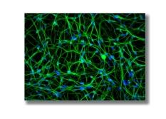
Figure 1 - HIP? Neurons differentiated to neuronal subtypes - After five weeks of differentiation, cultures show (A) an abundance of MAP2-positive neurons express the synaptic markerSynapsin, (B) Astrocytes marked by GFAP, (C) ChAT-positiveCholinergic neurons and (D) Vglut1-positive Astrocytes andGlutamatergic neurons. Nuclei are labeled with the Hoechst3342(blue). Scale bars= 200 microns.
Used as in vitro Models of Neural Disease
HIP? Neurons produce consistent, mature neuronal cultures for in vitro models of neural diseases. Through collaboration with SynAging SA [Nancy, France], HIP? Neurons (human induced pluripotent neurons ) have been used to create reliable in vitroassays to study the neurodegenerative properties of amyloid betaoligomers (AbO) and alpha-synuclein oligomers (aSO) that mimic Alzheimer’s and Parkinson’s Diseases, respectively.
View the press release here>
Alzheimer’s Disease Model
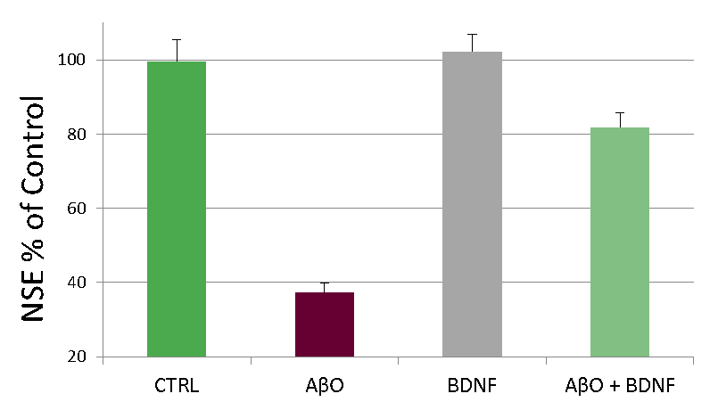
Figure 2. HIP? Neurons exhibit neurotoxicity in a model of Alzheimer’s Disease. Five-week old HIP? Neurons express neuronal and synaptic markers, including MAP2, PSD95 and synaptophysin (data not shown).After treatment for 24 hr with AbO, BDNF or AbO and BDNF, ELISA was performedfor neuron specific enolase (NSE), a specific marker for neurons. Data courtesy of SynAging.
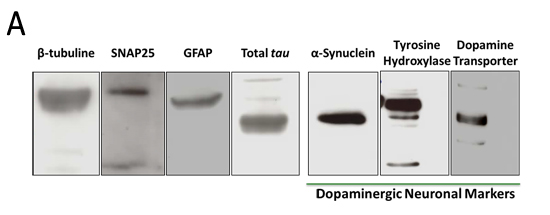
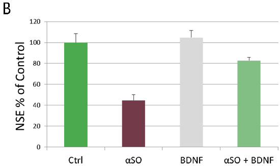
Figure 3. HIP? Neurons exhibit neurotoxicity in a model of Parkinson’s Disease. A) Differentiated HIP? Neurons express dopaminergic markers. B) HIP? Neurons cultured for five weeks and expressing dopaminergic markers were treated for 24 hr with alpha-synuclein oligomers(aSO), BDNF, or aSO and BDNF, ELISA was performed for neuron specific enolase(NSE), a specific marker for neurons.Data courtesy of SynAging. 
技術支持:易動力網絡